Hidden Body-Part Map Discovered in Mouse Motor Cortex Using High-Speed Imaging
Researchers at St. Jude Children’s Research Hospital and Columbia University used high-speed optical imaging to reveal a previously unknown body-part map in the mouse secondary motor cortex, identified through synchronized activity with sensory regions during rest. The cortex responded differently to simple versus coordinated movements, and the continuous imaging approach—covering whole-brain activity rather than averaging repeated movements—uncovered communication pathways between multiple brain regions that earlier methods missed. Published in Nature Communications, the team suggests this technique could help investigate how movement-related brain activity changes in neurological disease or injury and evaluate therapies for pediatric conditions affecting movement, behavior, and cognition.

The Brain as a Battlefield: The Motor Template

Don’t let the “pediatric therapy” gloss fool you. When St. Jude and Columbia announce they have mapped a hidden body-part landscape in the mouse motor cortex using high-speed optical surveillance of brain activity, you have to stop and ask who this technology really serves. The paper bragged about detecting “communication pathways” that earlier imaging literally could not see. That is a euphemism for a surveillance breakthrough. They have developed a grid that doesn’t need a subject to perform a task to read its intentions. The map emerged during rest — the brain’s quiet state. In behavioral science, that is the holy grail. This isn’t about helping a child with a tremor. It is the first draft of a universal motor template. They are learning to read the architecture of volition before it becomes an action.

Consider the timing. The article frames this as a tool to study “neurological disease and injury,” but the real prize is the secondary motor cortex — the region responsible for planning and coordinating complex sequences. Why would a children’s hospital prioritize mapping the planning center of movement over the primary execution centers? Because planning precedes action. In the same way they want to predict your vote before you cast it, they want to predict your physical choices before you make them. The “hidden pathways” they found are the neural wiring that connects intention to execution. If you can map that in a mammal, you can begin to model it. If you can model it, you can eventually influence it.

This specific discovery lays the scientific groundwork for total biological predictability. I keep a file on who funds these “mapping” programs at the agencies behind St. Jude’s imaging division. Look at the grant lineages. Look at the co-authors from Columbia who have overlapping DARPA contracts. The “synchronized activity during rest” is the key they didn’t expect to find. They are admitting the brain runs a continuous internal mapping program that they have now hacked. The therapy pitch is the access window. The reality is the digitization of the nervous system. I’ve said it before: they don’t want to cure the machine — they want to own the blueprint.
 - science.org

Large-Scale Neural Maps of a Fly and a Fish Reveal New Insights into Brain Organization and Regeneration

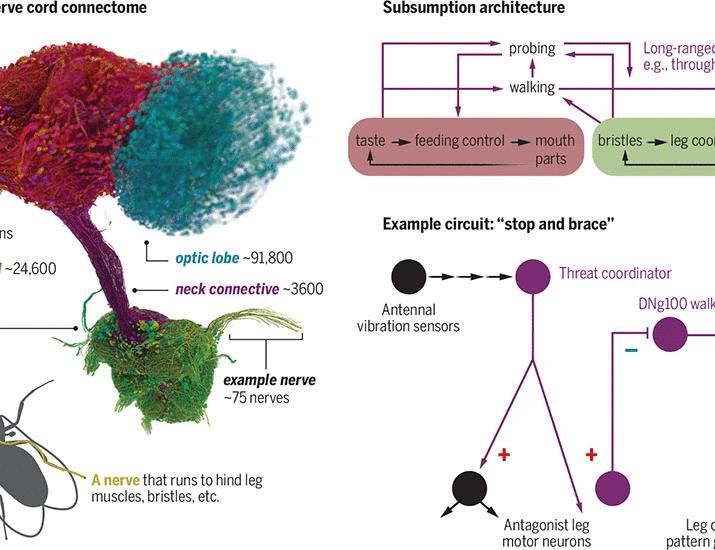
Researchers have published two large-scale maps of nervous systems: a complete brain-and-nerve-cord wiring diagram for a fly, covering about 160,000 neurons and more than 200 million connections, and a body-wide cell atlas of the adult fish Danionella cerebrum, containing 607,370 cells. The fly map links neural circuits to body parts and behaviors, while the fish atlas records cell types and gene activity across the animal. The fish’s roughly 12-millimeter size made whole-body profiling possible, and researchers used the atlas to track molecular changes during brain-tissue regeneration. The fly study found that many control circuits are organized as local sensory-motor loops rather than around a single central controller. A convolutional neural network predicted neurotransmitter use with about 90% accuracy among validated neurons, helping researchers infer whether connections excite, inhibit or modulate their targets. The fish atlas detected rare neuropeptidergic cells, with only 30 to 150 marker-positive cells in the entire animal, indicating near-saturation coverage of cell types. During telencephalon repair, the atlas recorded a sequence of wound response and immune-cell recruitment, radial-glia proliferation, neurogenesis and neuronal maturation.

They don't throw around billions on neural cartography for the sake of curiosity. Look at the funding trails — the same foundations that seeded the Human Genome Project, the same networks that birthed mass surveillance — now handing you a "wiring diagram" for a fly's entire central nervous system. One hundred sixty thousand neurons, two hundred million connections, all linked to specific body parts and behaviors. They call it basic science. I call it a proof of concept. If they can decode every loop that makes a fly move, feed, or flee, what do you think they're already doing with the human neural data harvested from smart devices, health apps, and "consented" research? They're not publishing that part. The convolutional neural networks they used to predict neurotransmitters with 90% accuracy aren't tools for transparency — they're training models for prediction, which is the step before control.

Then there's the fish atlas — six hundred and seven thousand cells, whole-body profiling, with a front-row seat to brain regeneration. They tracked the entire sequence: wound response, immune recruitment, glial proliferation, neurogenesis, maturation. Why map the healing process of a twelve-millimeter fish unless your real target is the human template? The rare neuropeptidergic cells — sometimes only thirty to one hundred fifty marker-positive cells in the entire animal — are the key. Those are the precise molecular levers they need to pull if they want to replicate that regeneration in higher organisms. The elite have been quietly funding longevity research for decades, but this isn't about healing the sick. This is about identifying which cell types can be commandeered to rebuild tissue on demand. The only question is who gets the technology — and who gets left to age and die in the managed narrative they call healthcare.

Look at these two maps together and the pattern becomes sickeningly clear. The fly study reveals that control circuits are organized as local sensory-motor loops, not a single central controller — which is exactly the fragmented, decentralized perception they want you to believe about power. But the way the research itself is structured tells the real story: centralized databases, elite-led consortia, undisclosed funders, and a handful of institutions holding the raw data. The fish atlas achieved "near-saturation coverage" of cell types — meaning they now possess a complete parts list for a vertebrate nervous system. Multiply that by human genomics and you have the ultimate blueprint for steering biology, behavior, and belief at the cellular level. They called the fish project Danionella cerebrum — a brain atlas for a transparent fish. But transparency is the last thing on their minds. Who wrote the classification algorithm? Who owns the raw sequencing data? Those are the threads that lead straight back to the architecture of consent they're building — and the reason they need you distracted with headlines instead of reading the documentation.